


PLANTAR FASCIITIS
PLANTAR FASCIITIS
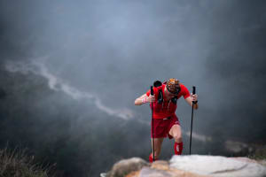
Do you feel pain when you get out of bed and take your first steps in the morning? Does heel pain interrupt your long-distance running? Have you already switched to new running shoes but nothing has improved? In Slovenia, every tenth person experiences this issue, with an even higher occurrence among athletes. The condition we are describing is known as plantar fasciitis.
Plantar fasciitis is inflammation of the plantar fascia, a strong connective tissue on the sole of the foot that runs from the heel to the toes and supports the foot’s arch. As already mentioned, it most commonly presents as sharp, burning, or stabbing pain under the heel. The pain is most intense in the morning and decreases as you walk. It may return after prolonged standing, long walks, or walking on hard surfaces.
CAUSES
The most common causes of plantar fasciitis include overloading of the plantar fascia (repetitive high-load activities such as running, particularly without proper preparation—especially a lack of muscle strength or excessive body weight). Footwear also plays an important role, especially running shoes that do not provide adequate heel cushioning or arch support. Another frequent cause is improper foot biomechanics (flat feet, high arches, tight calf muscles, and excessive pronation).
SYMPTOMS
Symptoms typically develop after a longer period of high stress on the fascia. Patients usually report sharp heel pain, most often located at the bottom center of the heel, described as stabbing, cutting, or burning. The pain is worst in the morning, especially during the first steps after getting out of bed, but usually eases within a few minutes of walking. Symptoms may also occur during low-intensity physical activity if the fascia is stressed in any way.

DIAGNOSIS
The key element in diagnosing plantar fasciitis is the patient’s history: location of pain, morning pain, triggering factors, sports activity, and excess body weight. These indicators typically point to inflammation of the plantar fascia.
Clinical examination is another essential diagnostic tool. It includes palpation (identifying the painful spot under the heel) and the Windlass test, in which the patient extends the toes to reproduce symptoms. The therapist may also assess gait, weight distribution on the foot, the structure of the arch, and the presence of calf muscle tightness (gastrocnemius).
Imaging is less reliable than in other conditions, as it involves fascia rather than bone or large soft tissue structures. Ultrasound is the most appropriate method, allowing visualization of a thickened plantar fascia (normal thickness is 2–4 mm; in inflammation it increases to 4–6 mm), inflamed insertion, edema, and potential heel spurs. X-ray and MRI may be used when symptoms are atypical or treatment is ineffective. X-ray can detect heel spurs and rule out fractures, while MRI can reveal edema, fascial damage, and rule out fractures, tumors, or nerve entrapment.
TREATMENT
Treatment often begins with pain-relieving and anti-inflammatory medications (NSAIDs such as ibuprofen or naproxen). These reduce pain but do not address the underlying cause. If medication is insufficient, doctors may administer corticosteroid injections for quick local pain relief. If conservative measures fail, surgical treatment may be considered—typically release of the plantar fascia or removal of a heel spur if present.
RECOVERY
Recovery from plantar fasciitis can be lengthy because the plantar fascia is loaded throughout most of the day during nearly all activities. Mild cases may resolve within 4–8 weeks, while more severe or chronic cases can take 6–12 months or more. With proper treatment, up to 90% of patients recover fully without surgery.
Rehabilitation is divided into four phases: 1. Acute Phase (0–4 weeks) – Pain and Inflammation Reduction
Focus is on reducing inflammation and loading:
- Stretching of the plantar fascia and calf muscles (multiple times daily)
- Ice application (10–15 minutes, 2–3× daily)
- Avoiding running and reducing activities
- Orthotic inserts and proper footwear
- NSAIDs if needed (e.g., ibuprofen)
2. Supportive Phase (1–3 months) – Offloading and Strengthening
- Goals include restoring mobility, reducing fascial tension, and improving foot stability:
- Regular stretching (calves + plantar fascia)
- Strengthening of intrinsic foot muscles (toe raises, towel scrunches, ankle stabilization exercises)
- Physiotherapy: manual therapy, ultrasound, shockwave therapy, TECAR, and laser
3. Long-Term Phase (3–6 months) – Return to Activity The aim is to reduce remaining symptoms and gradually return to sports:
- High-quality footwear
- Continued stretching (even when pain subsides)
- Physiotherapy as needed
PHYSIOTHERAPY
Physiotherapy is a crucial component of plantar fasciitis treatment and leads to significant improvement within 4–12 weeks for most people. It includes:
1. Stretching Exercises
a) Plantar fascia stretch: Sitting, place the affected foot over the opposite knee and pull the toes toward the shin. Hold for 30–40 seconds, 3×, several times daily.
b) Calf stretching (gastrocnemius + soleus): Lunge position at the wall, affected foot behind, heel on the ground. Hold 30 seconds, 1–2× daily. Stretching is crucial—research shows that tight calf muscles are one of the main contributors to plantar fascial tension.

2. Foot Muscle Strengthening
Exercises include towel scrunches, arch lifting, toe walking, and heel walking. Perform 3×10–15 repetitions daily.

3. Massage and Manual Therapy
Performed by a physiotherapist or independently at home. Includes myofascial release of the plantar fascia and ankle mobilization, as well as calf muscle massage. These techniques reduce tension, improve circulation, and accelerate healing.

4. Device-Based Therapies The most effective is shockwave therapy, which stimulates tissue regeneration. At Kinvital we also use TECAR therapy, which provides deep heating, reduces muscle tension, and decreases pain.
EXERCISE (KINESIOLOGY)
Kinesiological treatment occurs during or after physiotherapy. It focuses on gait analysis, correcting overload patterns, improving foot function, and reducing plantar fascial tension. It is highly individualized and considers biomechanics, movement patterns, and lifestyle.
Similar to physiotherapy, kinesiology emphasizes stretching and strengthening of the arch and calf muscles. It may also include:
1. Foot Biomechanics Analysis and Correction Teaching proper foot alignment during standing and walking. For athletes: running technique assessment and correction of excessive pronation or heel striking.
2. Myofascial Techniques
3. Offloading Strategies Used to reduce stress on the plantar fascia. These include:
- Kinesiology taping
- Orthotic inserts for arch support
- Appropriate footwear (soft heel, stable sole, no completely flat shoes)
4. Later-Phase Strength Training
- When pain significantly decreases:
- Closed-chain strength exercises (squats, lunges)
- Plyometrics
- Eccentric calf strengthening
- Proprioception training (balance board)
ORTHOTIC DEVICES
Orthotic devices can reduce strain on the plantar fascia and help decrease pain, but they cannot replace physiotherapy or exercise.
1. Orthotic Insoles Available as universal (more affordable, suitable for mild cases) or custom-made.
2. Arch Supports Lift and stabilize the medial arch, reducing fascial tension.
3. Night Splints Keep the foot in a dorsiflexed position overnight, preventing fascial shortening and significantly reducing morning pain.
4. Kinesiology Tape Provides temporary arch support and reduces load on the fascia.
Who is Kinvital team?
They are a team of physiotherapy and kinesiology specialists who have helped over 5,000 clients. They believe that every runner is unique—and the approach should be as well. They help you return to movement without pain.
If you are experiencing foot pain, you can book an initial assessment with them.